DDQX Learning · clinical-reasoning demonstration
When a smaller ventricle creates a bigger problem
A complex USMLE Step 1–style cardiovascular case demonstrating CPR Applications™ and the 5P Approach™.
One original teaching example · Select an answer for feedback · No score or learner data collected
In cardiovascular physiology, a severe obstruction does not always mean a small valve opening fixed in place. Sometimes the severity of an outflow obstruction changes from one moment to the next as loading conditions alter ventricular geometry. This case asks you to explain that change—and a second finding that appears alongside it.
01 · Your clinical challenge
An exertional episode with two competing murmurs
A 34-year-old man presents after nearly fainting while walking briskly uphill. He has experienced intermittent exertional breathlessness and chest discomfort for a year. Two days ago, he developed vomiting with poor oral intake. A clinician also started him on amlodipine for hypertension one week ago. His father died unexpectedly at age 42.
His supine blood pressure is 112/70 mm Hg and pulse is 94/min. A systolic crescendo–decrescendo murmur is heard along the left sternal border. Its intensity falls while he squats and increases when he returns to standing. An additional late-systolic apical component is heard.
Focused echocardiographic findings
- Basal interventricular septum: 21 mm
- Left ventricular cavity: relatively small; ejection fraction: 74%
- Aortic valve leaflets: normal morphology and mobility
- Peak Doppler LV outflow tract gradient: 18 mm Hg supine → 72 mm Hg during upright provocation
- A late-systolic, posteriorly directed mitral regurgitation jet becomes more prominent during provocation; no leaflet flail is seen
Which mechanism most directly accounts for both the increased outflow gradient and the accompanying mitral regurgitation?
Choose A–E below. Each option opens its own immediate explanation. Only one answer panel is intended to remain open at a time. This demonstration does not record or grade your selection.
02 · Build a spatial model
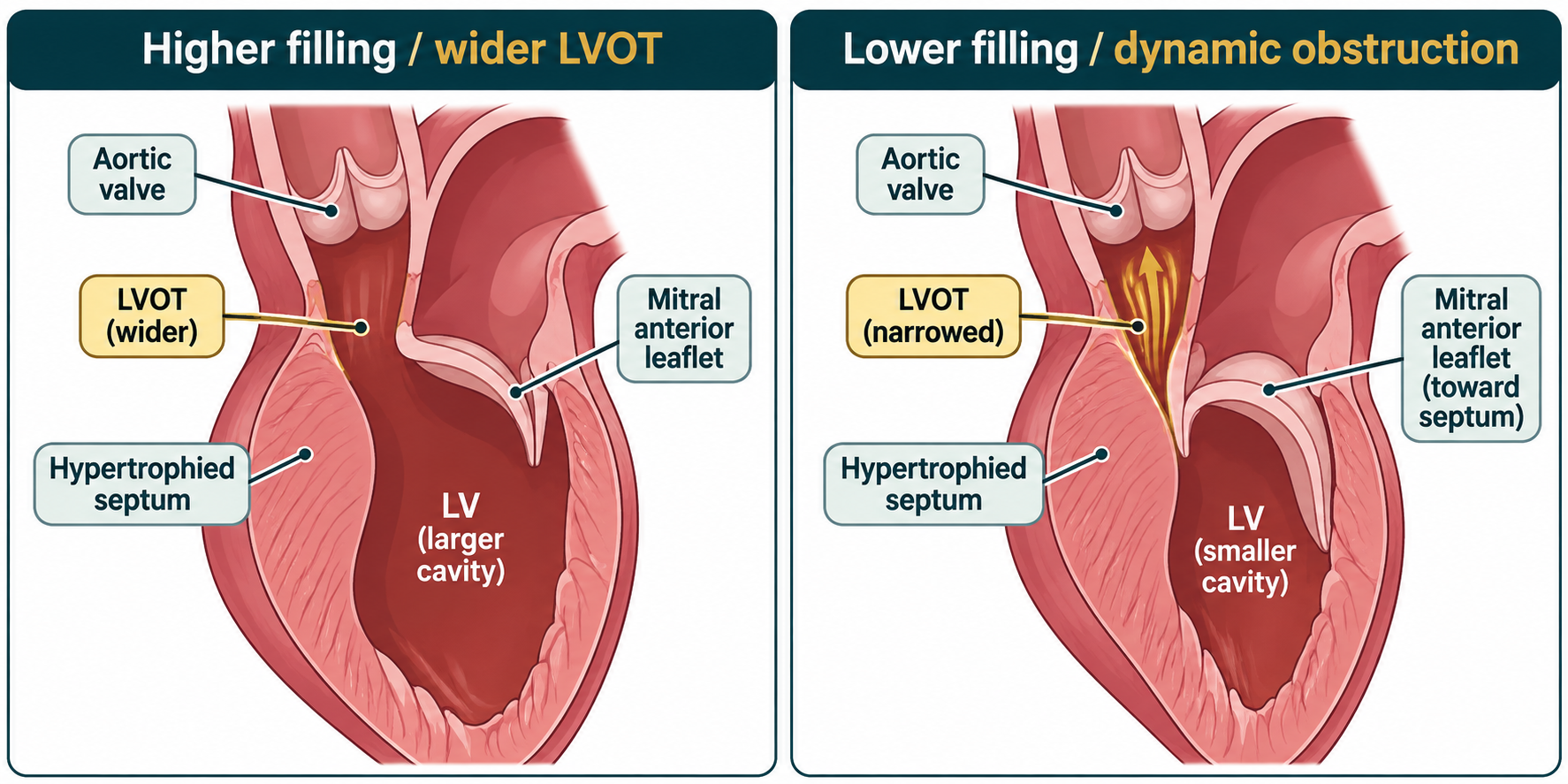
See what happens when filling falls
03 · Organize the knowledge
Concept → Pattern → Rule → Applications
CPR Applications™ organizes material you already encounter in physiology, pathology, and pharmacology into a chain that can be used on unfamiliar cases.
Concept · The mechanism
A normal left ventricle fills during diastole and ejects through the LV outflow tract and aortic valve. In hypertrophic cardiomyopathy (HCM), myocardial hypertrophy and impaired relaxation can reduce effective filling. In its obstructive form, basal septal thickening and abnormal mitral valve–flow interaction cause systolic anterior motion (SAM) of the anterior mitral leaflet toward the septum, dynamically reducing the outflow area.
That leaflet may also fail to meet its partner normally during systole, generating secondary mitral regurgitation—often directed posteriorly. The outflow gradient and the regurgitant jet may therefore arise from the same process.
A Step 1 trap: An ejection fraction of 74% describes the proportion of end-diastolic volume ejected. It does not establish normal filling, an adequate absolute stroke volume, or absence of severe dynamic obstruction.
Pattern · The discriminating evidence
Several findings become decisive when interpreted together: marked septal hypertrophy, a small vigorous LV, normal aortic leaflets, a murmur that strengthens with standing, a large provoked LVOT gradient, and late-systolic posteriorly directed regurgitation without leaflet flail.
The family history makes inherited HCM important to consider, but it is not the mechanism being asked for. The key discriminator is load-dependent subvalvular narrowing accompanied by SAM-related regurgitation, not a fixed aortic lesion or primary valve rupture.
Rule · Predict the direction of change
The inverse often holds: increasing filling can enlarge the cavity and reduce the gradient. Increasing afterload tends to lessen obstruction, whereas reducing afterload or increasing contractility can aggravate it. These are physiologic tendencies, not rules that predict every individual measurement.
Applications · Use the mechanism elsewhere
- Bedside physiology: Standing and Valsalva strain usually increase the dynamic murmur; squatting and handgrip often soften it through different loading effects.
- Pharmacology: Loss of circulating volume and arterial vasodilation can exacerbate the obstruction. Nonvasodilating beta-blockers are a common first-line option for symptomatic obstructive HCM, but this vignette asks about physiology rather than selecting treatment.
- Cardiac pathology: Septal hypertrophy and impaired relaxation can coexist with a preserved or high ejection fraction.
- Interpretation: A provocable gradient of 72 mm Hg is potentially clinically important, but presyncope and family history also call for an independent evaluation of arrhythmic and sudden-death risk.
Compare the direction of the maneuvers
| Maneuver | Principal change | Typical obstructive HCM response |
|---|---|---|
| Stand from squat | Less preload and lower afterload | Greater obstruction / louder murmur |
| Sustained squat | More preload and afterload | Less obstruction / softer murmur |
| Valsalva strain | Less venous return | Greater obstruction / louder murmur |
| Isometric handgrip | More systemic afterload | Less obstruction / softer murmur |
Compare mechanisms rather than memorizing four isolated sound changes. Real Doppler gradients and murmur intensity also depend on the patient’s loading conditions and how the maneuver is performed.
04 · Solve the problem deliberately
The 5P Approach™ walkthrough
The CPR framework provides the organized knowledge. The 5P Approach™ structures what the learner does with it while answering and reviewing the item.
1 · Prioritize
Keep the evidence that discriminates mechanisms: 21-mm basal septum; small hyperdynamic LV; anatomically normal aortic valve; obstruction rising 18 → 72 mm Hg on standing; and a posteriorly directed, late-systolic mitral jet without flail. Volume loss and vasodilation make the loading changes plausible. The patient’s family history is important clinically but does not, on its own, answer the mechanism question.
2 · Paraphrase
“A smaller, thick-walled LV becomes more obstructed on standing, and mitral regurgitation increases at the same time. What single mechanical event explains both changes?”
3 · Prognose
Before looking at A–E, predict: “Lower venous return makes the cavity smaller, bringing the mitral leaflet toward the enlarged septum during systole. This narrows the outflow tract and prevents normal mitral coaptation.” The expected answer must connect both the outflow gradient and regurgitation.
4 · Pick
B matches the predicted mechanism. A attributes the physiology to a fixed aortic lesion, C reverses the contractility effect, D reverses the usual afterload effect, and E mistakes a possible consequence (mitral regurgitation) for a primary structural injury. Do not choose an answer just because it mentions a real disease that can produce a systolic murmur.
5 · Post-Mortem
Identify the variable manipulated (ventricular filling), the anatomical mediator (leaflet–septal interaction), and the observed outputs (higher gradient plus secondary regurgitation). Then ask what would change with the opposite loading intervention, or what evidence would instead support primary mitral disease. That analysis matters more than remembering the answer letter.
05 · Illustrative reasoning error
When a true clue becomes the wrong conclusion
Hypothetical learner selects E: “The patient has mitral regurgitation. A ruptured papillary muscle can cause a systolic murmur and severe regurgitation, so the mitral valve must be the source of the obstruction.”
What the learner noticed: A real pathologic finding—the regurgitant jet.
Where the inference breaks: The learner treats the jet as the initiating lesion, without checking whether a mechanism already explains it as a downstream consequence. There is no infarction pattern or flail leaflet, while the provoked outflow obstruction and posterior jet fit systolic anterior motion.
The corrected decision: If two abnormalities appear together, ask whether one mechanism can produce both. Distinguish primary mitral injury from mitral regurgitation secondary to dynamic LV outflow obstruction.
06 · Targeted remediation
The feedback is a next action
Reasoning vulnerability: Recognizing an associated finding but failing to determine whether it is a cause or an effect.
Repair task: Draw two arrows from reduced LV filling: one toward the outflow gradient, another toward mitral coaptation. Explain why both arrows pass through systolic anterior motion of the mitral valve.
Control comparison: Put that chain beside the mechanism of papillary muscle rupture after infarction. In that contrasting condition, primary loss of valve support—not dynamic leaflet–septal interaction—initiates the regurgitation.
Success criterion: Without a maneuver table, correctly predict the direction of change in obstruction when preload or afterload rises, and state why a preserved ejection fraction does not exclude low effective forward output.
Check your transfer
Suppose the patient’s resting gradient is low again, but a repeat test demonstrates substantial mitral regurgitation without systolic anterior motion or a provoked LV outflow gradient. Should that regurgitation automatically be attributed to obstructive HCM?
Open the transfer explanation
No. HCM and primary mitral valve disease can coexist. If the causal pathway through SAM is not present, investigate the mitral valve and subvalvular apparatus rather than automatically labeling the jet as obstruction-related. A transferable mechanism is useful partly because it tells you when your original explanation may no longer hold.
The purpose of the demonstration
CPR Applications™ turns cardiovascular facts into a coherent mechanism: Concept → Pattern → Rule → Applications. The 5P Approach™ turns that mechanism into a repeatable process for solving questions and learning from errors.
This page is one public teaching example. DDQX’s separate CPR Applications digital product is still in development; this example does not represent its released assessment, adaptive feedback, or learner tracking.
Explore CPR Applications™ · Learn the 5P Approach™ · Step 1 resources
Sources and scope
DDQX Learning, CPR Applications for USMLE Step 1, Chapter 6 (cardiac loading conditions and §6.26, hypertrophic cardiomyopathy); 2024 AHA/ACC/AMSSM/HRS/PACES/SCMR Guideline for Hypertrophic Cardiomyopathy; Merck Manual Professional: Hypertrophic Cardiomyopathy; and Merck Manual: Maneuvers That Aid in Diagnosis of Murmurs.
This original fictional vignette and its illustration are for medical education; they are not clinical instructions, a diagnostic tool, or official USMLE/NBME materials. Symptoms such as exertional near-syncope and a family history of sudden death require patient-specific clinical evaluation.